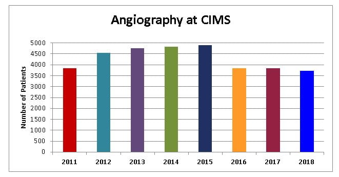
Angiography
Angiography is the process of taking pictures of the inside of the heart or blood vessels. The picture itself is called an angiogram. The angiogram allows your doctor to check the inside of a blood vessel to see if it is narrowed or blocked.
A special dye that shows up in an x-ray is injected into a blood vessel through a needle or a small tube called a catheter. As the dye flows in the blood vessel, an x-ray machine rapidly takes a series of pictures. Sometimes the pictures are taken so fast that they form a movie of the progress of the dye. Cardiac catheterization and the injection of dye into the arteries is the best way to study the coronary arteries.
Chest X-Ray
X-rays are a form of electromagnetic energy, or radiation. X-rays are able to penetrate body tissues. They are used to create pictures of body structures on film. An x-ray of your chest can show:
X-rays are a form of electromagnetic energy, or radiation. X-rays are able to penetrate body tissues. They are used to create pictures of body structures on film. An x-ray of your chest can show:
if the heart is enlarged or normal signs of heart failure and fluid overload pneumonia or a collapsed lung tumors in the lung that could mean cancer.


